Screening Applications & Diagnostics
Poster Session A
(1020-A) AI-powered Single Cell Drug Screening in Nanowell Plates
Tuesday, May 28, 2024
16:30 - 17:15 CEST
Location: Exhibit Hall

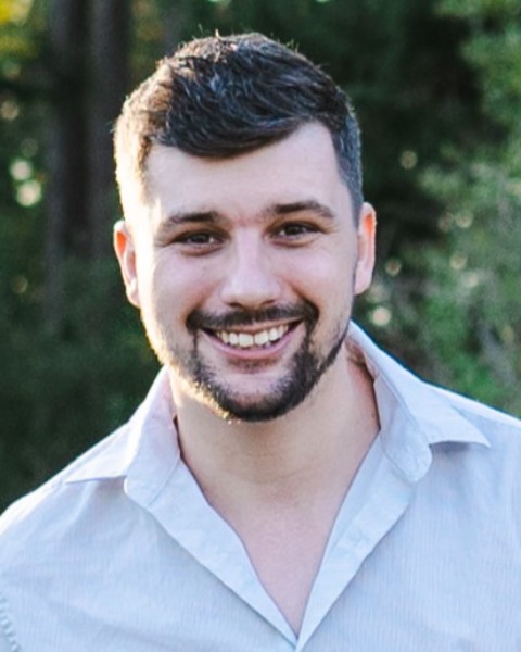
Samuel G. Berryman, n/a
CEO, Graduate Student
ImageCyte, University of British Columbia
Vancouver, British Columbia, Canada
Poster Presenter(s)
Abstract: Conventional drug-screening workflows typically involve depositing cell samples into microwell plates to treat them using novel compounds, and then subsequently, measure their response using microscopy. Recently, emerging AI tools presented new approaches for analyzing these microscopy images to extract additional information, such as cell state, function, and phenotype. However, a major challenge in these workflows is preparing standardized images of single-cells that can be used in the training of machine learning models. This difficulty arises from the fact that individual cells are difficult to segment, and are often confounded by the presence of neighbouring cells. For this reason, it would be desirable to physically separate the cells so that they can be more easily analyzed.
In response to this need, we developed glass-bottom nanowell-in-microwells plates, which subdivide standard microwells into thousands of nanowells. The nanowell microstructures are fabricated using photolithography, which enable a high-density number of nanowells on a glass bottom substrate. The nanowells can be as small as 25 x 25 µm, allowing approximately 2.5 million nanowells to fit on a single 384-well plate. The nanowell microstructures are also black in color, which suppresses background fluorescence and provide high-contrast between the nanowell walls and the glass-bottom substrate to simplify the extraction of single-cell images.
We performed two studies to demonstrate the potential utility of these nanowell-in-microwell plates for AI-powered drug screening. In the first study, we exposed MDA-MB-231 cells to 20 anti-cancer compounds with 10 different mechanisms-of-action. The cells were treated with varying concentrations of each compound and imaged using an inverted microscope. We used these images to train a machine learning model to classify cell images based on mechanisms-of-action. Our study demonstrates the potential to predict the mechanism-of-action of novel compounds, to assess the value of potential hits.
In the second study, we again exposed MDA-MB-231 cells to varying concentrations of anti-cancer compounds. We acquired brightfield images of each cell, as well as their viability using a live/dead stain. We then used this dataset to train a machine learning model to determine cell viability from the brightfield images alone. Finally, we used the machine learning model to determine the viability of previously unseen cells, acquired at varying drug concentrations, to construct dose-response curves and obtain IC50 values. Our results demonstrate the potential to perform stain-free drug screening to reduce consumable needs and increase screening throughput.
In summary, we show that nanowell-in-microwell plates provide a simple method to acquire large data sets of standardized images for cell morphology analysis and machine learning. This approach enables the use of sophisticated AI tools to improve value of the microscopy image datasets acquired during high-throughput screening.
