Frontiers in Technology
Poster Session B
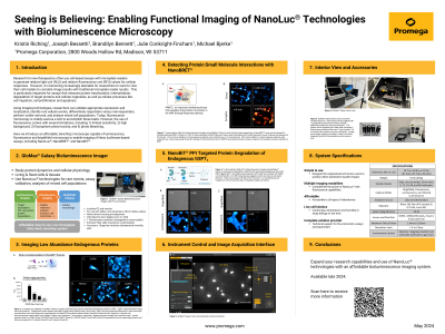
(1151-B) Seeing is Believing: Enabling Functional Imaging of NanoLuc® Technologies with Bioluminescence Microscopy
Wednesday, May 29, 2024
10:30 - 11:15 CEST
Location: Exhibit Hall


Julie J. Conkright-Fincham, PhD (she/her/hers)
Strategic Collaborations Manager
Promega Corporation -
Madison, WI, United States
Poster Presenter(s)
Abstract: Research for new therapeutics often use cell-based assays with microplate readers to generate relative light unit (RLU) and relative fluorescence unit (RFU) values for cellular responses. However, it is becoming increasingly desirable for researchers to want to view their cell models to correlate image results with traditional microplate reader results. This is particularly important for assays that measure protein translocation, internalization, degradation of target proteins and cellular organelles, as well as cellular processes like cell migration, cell proliferation and apoptosis.
Using imaging technologies, researchers can validate appropriate expression and localization, identify rare cellular events, differentiate responders versus non-responders, perform outlier removal, and analyze mixed cell populations. Today, fluorescence microscopy is widely used as a tool to accomplish these tasks. However, the use of fluorescence comes with several limitations, including 1) limited sensitivity, 2) high background, 3) fluorophore photo-toxicity, and 4) photo-bleaching.
Here we introduce an affordable, benchtop microscope capable of luminescence, fluorescence and brightfield microscopy to enable imaging of Nano luciferase-based assays, including NanoLuc®, NanoBRET™ and NanoBiT®.
