Frontiers in Technology
Poster Session B
(1143-B) Nuclei Segmentation on Brightfield Images using a pre-trained Artificial Intelligence (AI) Model
Wednesday, May 29, 2024
10:30 - 11:15 CEST
Location: Exhibit Hall

.jpg)
Scott Charles Cribbes
Market Development Leader
Revvity
GLASGOW, Scotland, United Kingdom
Poster Presenter(s)
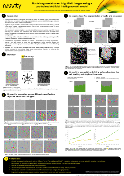
Abstract: In recent years, AI and machine learning-based image analysis processing has gained traction due to its promise to enable tasks that were not possible before, such as segmentation of nuclei on brightfield images. Brightfield images are rich in information but clearly lack the contrast and without specific staining presence in fluorescent images, making segmentation more challenging. Our findings show that a pre-trained model can segment nuclei on brightfield images without additional training input by the user that is typically required. This was achieved by developing an algorithm from several thousands of HCS generated images acquired in several cell lines with different magnification lenses to derive a universal deep learning model, all within the GUI. The advantage of using the pre-trained models is users don’t need to invest un-necessary time, concerns for computational power for the AI training process, or validation before analyzing the data. One area of applications that clearly benefits from brightfield segmentation is live cell imaging. AI based nuclei segmentation alleviates the need of a nuclei fluorescent dye, i.e., DAPI, Hoechst, or others used in traditional segmentation process, which are known to produce cytotoxicity and/or phototoxicity in live cells and limits longitudinal kinetic study experiments. Label-free brightfield segmented images completely abolishes all these concerns. We demonstrate that brightfield segmentation is ideal for cell tracking applications since it allows for time series image acquisitioning with high temporal resolution and minimized perturbation of the cells. Furthermore, the same data can be used to generate an AI-based digital phase image which is more robust against potential imaging artifacts than conventional digital phase images. Thereby, the cells can be segmented into nuclei and cytoplasmic compartments, or as a whole cell region without any fluorescent labels. Subsequently, all common “classical” analysis tools can be used for feature extraction, or any additional fluorescent channel acquired, including fluorescent proteins often used in live cell monitoring. In addition to live cell applications, brightfield segmentation can be applied to fixed samples freeing up a channel normally used for nuclei staining to allow expansion of target specific staining to increase multiplexing capabilities.
