Head of Cell Culture Application Lab Synentec Elmshorn, Schleswig-Holstein, Germany
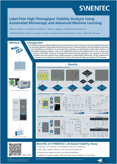
Abstract: Cell viability assays play a crucial role in various research fields such as drug discovery, cancer research, bioprocessing, and cell line development. The Trypan Blue (TB) assay is widely used in both academia and industry to assess cell viability. This dye exclusion assay enables the determination of cellular health by visually examining under a light microscope whether the cells incorporate or exclude the dye. However, TB has several limitations, including cytotoxicity at high concentrations or prolonged exposure, and it is classified as a carcinogen. Safe handling and disposal, particularly when mixed with genetically modified organisms, are complex due to the regulatory environment worldwide. As a result, there is a growing demand for reliable, efficient, and cost-effective alternatives that can overcome these challenges.
Our goal was to develop a solution that uses brightfield images of unlabeled cells captured with our widefield imagers to evaluate the viability of suspension cells using advanced machine learning (ML)/artificial intelligence (AI) algorithms. We created training data by staining a mixed population of live and dead CHO-K1 or HEK293T cells with Hoechst 33342, Calcein-AM, and Propidium Iodide (PI), and then imaged the cells using our automated high-content imager, CELLAVISTA® 4K. Based on this staining, we labeled the cells in the brightfield channel and classified them as live (Calcein-AM positive/PI negative) or dead (PI positive) using our proprietary software, AI-STUDIO+. The AI models were trained on this data to detect cells and assess their viability based on morphological differences. We confirmed the accuracy of the AI model by comparing its results with those obtained from traditional image processing of TB staining. Both assays provided similar results in terms of viability and total cell count, with the AI-analyzed data showing a lower standard deviation than the traditional TB assay. The AI model is integrated into our YT-SOFTWARE®, allowing imaging and analysis to be conducted within the same software ecosystem, thereby reducing time and costs for additional training and support.
In conclusion, we have developed a reliable tool for high-throughput automated evaluation of suspension cell viability, eliminating the need for toxic dyes and incubation steps.