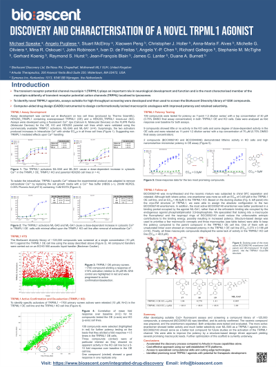
Senior Scientist/Team Lead BioAscent Discovery MOTHERWELL, Scotland, United Kingdom
Abstract: The transient receptor potential channel mucolipin 1 (TRPML1) is the most characterised member of the mucolipin subfamily of transient receptor potential cation channels (TRPML) localised to lysosomes, which also includes TRPML2 and TRPML3. TRPML1 is a non-selective cation channel permeable to Ca2+, Fe2+ and Zn2+, with its primary function being to induce Ca2+ release from endolysosomal compartments (1,2). TRPML1 channels are expressed ubiquitously, including in all regions of the brain. It plays an important role in neurological development and function as the loss-of-function of TRPML1 due to mutations in the human TRPML1 gene, MCOLN1, causes the rare neurodegenerative lysosomal storage disorder (LSD) mucolipidosis type IV (MLIV), characterised by intellectual disability, psychomotor retardation, hypotonia, cornea opacity and retinal degeneration (3). TRPML1-mediated Ca2+ release is also impaired in other LSDs, such as Niemann-Pick disease type C (NPC) (4). Activation of this channel is also linked to autophagic clearance of Aβ and tau in Alzheimer’s disease (AD) through the activation of transcription factor EB (TFEB) (5), leading to the suggestion that pharmaceutical targeting of TRPML1 could be an effective treatment option for the disease. To further investigate its therapeutic validity, the identification of novel, selective TRPML1 agonists would complement the recent discovery and characterisation of TRPML1 antagonists (6,7). To this aim we screened the BioAscent diversity library of ~125,000 compounds at 10µM in a TRMPL1 over-expressing (OE) HEK293 cell line using a FLIPR calcium mobilisation assay. Compounds were identified as hits if they were confirmed as showing ≥15% the agonistic effect of ML-SA5 but did not exhibit Ca2+ release in a TRPML1 knockout (KO) control HEK293 cell line. 109 compounds identified as active in the primary screen were selected for full concentration-response potency testing. Among them, Compound 1 (racemic) was shown to have an pEC50 of 6.2 ± 0.1 (n = 16) in the TRPML1 OE cell line, but no activity in the TRPML1 KO cell line (Fig. 2). This indicates that the calcium mobilisation observed in the TRPML1 OE cell line by 1 is mediated by selective activation of the TRPML1 channels. In comparison, ML-SA5 showed an pEC50 of 6.14 ± 0.16 (n = 10) in the TRPML1 OE cell line. Surprisingly, ML-SA5 exhibited Ca2+ release with a similar level of potency, pEC50 = 6.04 ± 0.086 (n = 5) in the TRPML1 KO cell line, albeit with a lower Emax.
References 1. Kiselyov K, Colletti GA, et al. Cell Calcium. 2011;50:288. 2. Di Paola S, Scottoo-Rosato A, Medina DL. Cell Calcium. 2018;69:112. 3. Bargal R, Avidan N, Ben-Asher E, et al. Hum Mutat. 2001;17:397. 4. Lloyd-Evans E, Morgan AJ, He X, et al. Nat Med. 2008;14:1247. 5. Medina DL, Di Paola S, Peluso I, et al. Nat Cell Biol. 2015;17:288. 6. Kriegler K, Leser C, et al. Arch Pharm. 2022;355:e2100362. 7. Leser M, Keller S, et al. Eur J Med Chem. 2021;210, 112966.

 , MRes photo")