European Application Engineers Hamamatsu Photonics Massy, Ile-de-France, France
Abstract: In drug discovery and development, there is a rapidly growing need to use physiologically-relevant cell culture models that are use of patient-derived and iPSC-derived disease modelling cells and/or 2D heterogeneous and 3D cell cultures. To advance fluorescent phenotypic assays and screenings using these cell culture models in microplates.
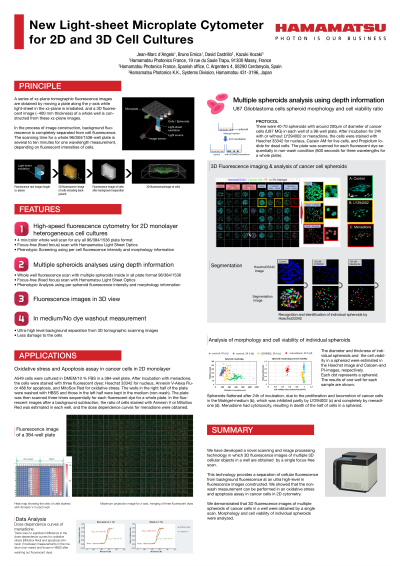
We have developed a new scanning and image processing technology, Zyncscan, in which a series of xz-plane tomographic fluorescence images are obtained by moving a plate along the y-axis while light-sheet in the xz-plane is irradiated, and a 3D fluorescent image of a whole well is constructed from these xz-plane images. This new scanning and image processing enables us to obtain 3D fluorescence images of multiple 3D cellular objects with various sizes and morphologies in a well by a single focus-free scan for a whole plate (no need to focus on each cellular object and to take multiple xy-plane images of the object). Additionally, fluorescence measurements and imaging can be performed in cell culture mediums containing serum and fluorescent dyes due to the complete separation of cell and background fluorescence.
We constructed new Light-sheet microplate cytometer for 2D and 3D cell cultures call CYTOQUBE using Zyncscan technology. We performed live-cell experiments to confirm and demonstrate the new functions mentioned.