Field Application Specialist Sun Bioscience SA Lausanne, Vaud, Switzerland
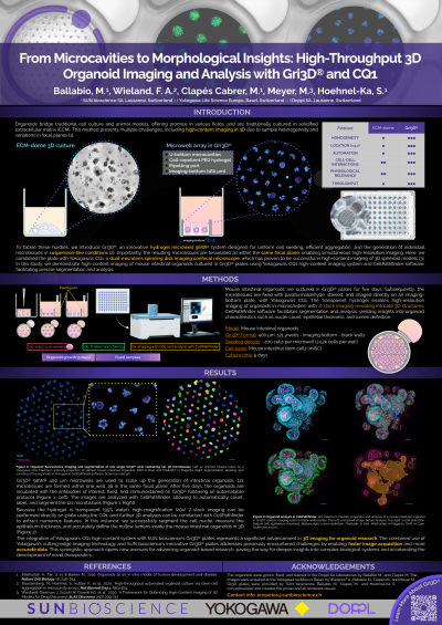
Abstract: Organoids bridge traditional cell culture and animal models, offering promise in various fields, and are traditionally cultured in solidified extracellular matrix. This method present challenges for high-content imaging in 3D due to sample heterogeneity and variations in focal planes. To address this, we present an innovative imaging approach integrating Yokogawa's CQ1 high-content imaging system with SUN bioscience's Gri3D® microwell plates. Our study demonstrates high-content imaging of mouse intestinal organoids cultured in Gri3D® plates using Yokogawa’s CQ1 and CellPathfinder software, facilitating precise segmentation and analysis.
Mouse intestinal organoids were cultured in Gri3D® plates, fixed, stained, and imaged using Yokogawa’s CQ1. The transparent hydrogel enabled high-resolution imaging of organoids in microcavities, with Z-stack imaging revealing intricate 3D structures. CellPathfinder software facilitated segmentation and analysis, yielding valuable insights into organoid characteristics such as nuclei count, epithelial thickness and lumen detection.
The integration of Yokogawa's CQ1 with SUN Bioscience's Gri3D® plates represents a significant advancement in organoid imaging, enabling faster image acquisition and more accurate data.

 photo")