Frontiers in Technology
Poster Session A
(1000-A) Design and validation of SPAchip® microdevices for continuous intracellular monitoring Reactive Oxygen Species (ROS) and molecular oxygen (O2) in living single cells.
Tuesday, May 28, 2024
16:30 - 17:15 CEST
Location: Exhibit Hall


David Garcia Soriano, A4CELL (he/him/his)
Chemical Specialist
A4CELL
Madrid, Madrid, Spain
Tony B. Poster Author(s)
Abstract: Oxidative metabolism and redox reactions are vital for living organisms. Last years, many researchers have studied the mitochondrial activity in the cell cycle that generates a small amount of highly reactive radicals known as reactive oxygen species (ROS), which often play an important role in cell signaling and defense against pathogens.[1] However, when the cell's antioxidant systems fail, or ROS production is increased due to failures in the mitochondria and the redox balance is chronically disrupted, a situation known as oxidative stress is generated in which numerous cellular components can be irreversibly damaged. This condition has been implicated in the pathogenesis of numerous disorders and diseases, such as diabetes, neurodegeneration, or cancer.[2]
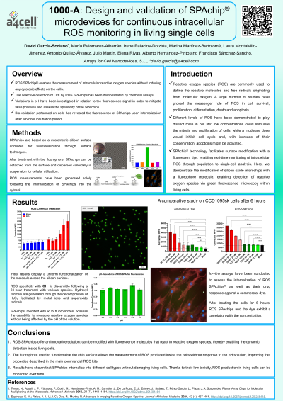
The presented technology introduces an innovative biosensor that enables measure the intracellular oxygen and detect the production of ROS within live cells through fluorescence microscopy. This lab-in-a-cell device is based on an inert 9-um2 silicon square functionalized with selected fluorescent probes. These Suspended-Planar Array chips (SPAchip®)[3] are passively internalized into the cytosol without causing cytotoxicity and allowing long-term cellular studies. Their potential to act as a unique cellular analysis tool enables multi-day monitoring of oxygen consumption rates (OCR) or ROS generated in any living cell type, from adherent tumor cells to heterogeneous and complex 3D cell models.
Cells are cultured with SPAchips inside and subjected to hypoxia conditions to evaluate oxygen consumption by treatment with respiratory chain uncouplers (Antimycin A, Rotenone, FCCP...), or ROS pulses by generation of superoxide anion (O2-·), hydrogen peroxide (H2O2) in the presence of Fe2+ for the generation of hydroxyl radicals (OH·) and shortwave light (405 nm laser) to generate metabolic stress. The response of the chips will be analysed by fluorescence microscopy and flow cytometry.
