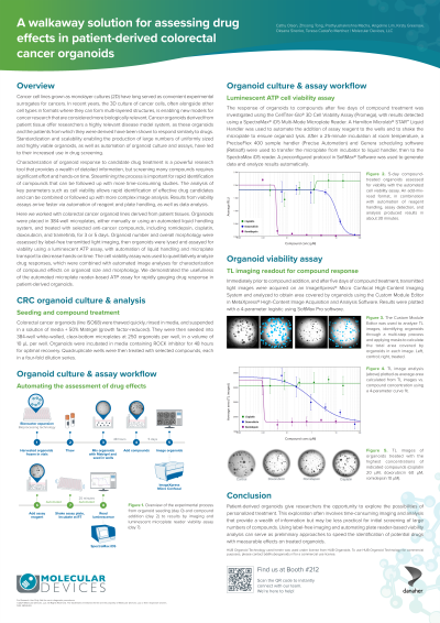
European Field Application Scientist Molecular Devices Wokingham, England, United Kingdom
Abstract: Cancer cell lines grown as monolayer cultures (2D) have long served as convenient experimental surrogates for cancers. In recent years, the 3D culture of cancer cells, often alongside other cell types in formats where they can form multi-layered structures, is enabling new models for cancer research that are considered more biologically relevant. Cancer organoids derived from patient tissue offer researchers a highly relevant disease model system, as these organoids and the patients from which they were derived have been shown to respond similarly to drugs. Standardization and scalability enabling the production of large numbers of uniformly sized and highly viable organoids, as well as automation of organoid culture and assays, have led to their increased use in drug screening.
Characterization of organoid response to candidate drug treatment is a powerful research tool that provides a wealth of detailed information, but screening many compounds requires significant effort and hands-on time. Streamlining the process is important for rapid identification of compounds that can be followed up with more time-consuming studies. The analysis of key parameters such as cell viability allows rapid identification of effective drug candidates and can be combined or followed up with more complex image analysis. Results from viability assays arrive faster via automation of reagent and plate handling, as well as data analysis.
Here we worked with colorectal cancer organoid lines derived from patient tissues. Organoids were placed in 384-well microplates, either manually or using an automated liquid handling system, and treated with selected anti-cancer compounds, including romidepsin, cisplatin, doxorubicin, and trametinib, for 3 or 5 days. Organoid number and overall morphology were assessed by label-free transmitted light imaging, then organoids were lysed and assayed for viability using a luminescent ATP assay, with automation of liquid handling and microplate transport to decrease hands-on time. The cell viability assay was used to quantitatively analyze drug responses, which were combined with automated image analyses for characterization of compound effects on organoid size and morphology. We demonstrated the usefulness of the automated microplate reader-based ATP assay for rapidly gauging drug response in patient-derived organoids.

 photo")