Senior R&D Manager Revvity Health Sciences, Inc. Lawrence, MA, United States
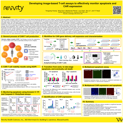
Abstract: Chimeric antigen receptor (CAR)-T cell therapy is one of the novel cellular therapeutic options for cancer patients, such as the B-cell malignancies. To effectively develop and manufacture CAR-T products, it is imperative to identify and assess the critical quality attributes (CQAs) of CAR-T cells. Recently, the Food and Drug Administration (FDA) has published the chemistry manufacturing and controls (CMC) guidance for human gene therapy. Cell concentration, cell viability, and CAR expression are important quality attributes that should be reliably measured during the entire process. In this work, we developed an image-based method to quickly count viable T cell, measure viability, and assess CAR expression. Using this new method, we compare different gene delivery procedures to make CAR T cells, mostly through the electroporation process. Our results showed that high percentage of apoptotic cells were introduced during the electroporation process, regardless of the CAR vectors. Cell viabilities were checked daily with AO/PI dual fluorescent staining method and the viability data were correlated with apoptosis data. On Day 1 after transduction, measured cell viabilities of transduced SupT1 cell samples decreased to ~60% - ~70%. Compared to measured cell viabilities of un-transduced and electroporation SupT1 controls of over 90%, it was identified that the introduction of plasmids, other than the electroporation process itself, induced 20 – 30% additional cell death. Our apoptosis data further confirmed that most of the cell death after electroporation were potentially due to cell apoptosis. The transduced SupT1 cell samples were able to recover as their viabilities increased to >90% on Day 3. Percentages of CAR expression in transduced SupT1 cell samples were characterized by staining with APC-conjugated specific anti-CAR antibody. As for the percentages of CAR expression measurements, we observed similar CAR expression results in different transduced SupT1 cell samples on Day 1 and Day 2. We confirmed the percentages of CAR expression results using the flow cytometer and did not see significant differences between Cellaca® PLX Image Cytometer and flow cytometer results. With the advantages of ease of use, visual verification with captured cell images, and higher-throughput capability, Cellaca® PLX Image Cytometer may be potentially used as the convenient benchtop system for rapid assessment of the quantity and quality of CAR-T cells, which may ultimately improve the productivity of development and manufacture of CAR-T products.